
A Critique of http://www.patspeer.com/
Chapters 18a, 18b, and 19b
It ain't what you don't know that gets you into trouble. It's what you know for sure that just ain't so.
~Mark Twain
Note 2: For my Dallas lecture (2009), see The JFK Skull X-rays: Evidence for Forgery
Note 3: ARRB summaries (Horne's Appendices 43-46) of the three forensic experts
Introduction to this Critique
Jim DiEugenio brought the extraordinary work of Pat Speer to my attention. Since Jim wanted my feedback, and because Speer's interests overlapped mine, I devoted several slides in my Dallas talk to Pat's two chief proposals: explanations for the 6.5 mm object on JFK's AP skull X-ray, and for the White Patch on the lateral skull X-rays. I first met Pat in the hallway after my talk, where I identified him by his name tag and we had a brief and courteous chat. I recall being surprised that he had not attended my lecture, although I later learned (from his website) that he had caught the last few minutes. It is the only time we have exchanged any dialogue. About a year later I visited his website again; that visit has prompted this review. At the above website, Speer has established a new record by nominating over 30 individuals for a rogue's gallery, i.e., individuals who have made (meaningful) mistakes in this JFK case:
| Aguilar | Horne | Myers (aka Meyers) |
| Baden | Kurtz | Morgan |
| Bell | Lattimer | Peters |
| Bugliosi | Lifton | Piziali |
| Crenshaw | Lindenberg | Robertson |
| Davis | Mantik | Spitz |
| Durnavitch | McAdams | Sturdivan |
| Fackler | McCarthy | Wecht |
| Fetzer | McClelland | White |
| Groden | McDonnel | |
After Speer's self-assured omniscience at ferreting out these blunders (and their guilty sponsors), I was not too daunted at seeing my own name among such an illustrious throng. However, it quickly got worse: Speer had nominated me for a special citation. Not only had I made (many) mistakes, but I had lied:
So, after seven decades, I finally qualified as a liar. Curiously, my problem has always been the reverse – that of being too honest. (Speer cites me as lying about the location of the (presumed) lead smudge on the Harper fragment and about the explanation for the White Patch.) My devout Pentecostal mother, who had persistently drummed one lesson into my childhood head – never to lie – would have risen from her grave had she heard that charge. I have never been able to shake those shackles (of never lying), and my children are afflicted as well. But Speer still wasn’t done – he gamely went on to proffer some other attention-grabbing remarks:
Before I began this project I knew virtually nothing about x-rays.
Durnavitch, and just about everybody else who's written about the x-rays, was wrong.
And yet it seems I've uncovered many issues not addressed by the so-called experts.
I offer one important clarification in this critique. After my Dallas lecture I recognized, with some regret, that I had left the audience with a confused picture of the (apparent) site of lead debris on the Harper fragment. Speer gets credit for also noticing this, and the audience has my apologies. The confusion arose from new evidence on the Harper X-ray, discovered by John Hunt. The X-ray showed the metal debris to lie at the opposite pole of the Harper fragment from where I had originally placed it (a placement that had been based solely on the photographs). For my Dallas lecture I showed only a close-up image (slide 19) of the Harper X-ray (courtesy of John Hunt), but I should have shown the entire X-ray. I correct that oversight here. However, if this new site for metal is accepted, Speer's placement of the Harper fragment (like Riley's and Angel's) suffers grievous trauma.
Chapter 18: X-ray Specs
Note 5: These twenty questions were prompted by Speer's comments, although the wording here is (mostly) my own.
No, it does not matter. The autopsy suite had no installed unit, so the only option was a portable unit. But Speer quotes (p. 7) Dr. John Ebersole (the autopsy radiologist, who practiced as a radiation oncologist): for the evening's chief purpose (locating metallic debris), this unit was quite satisfactory. I agree that a permanently installed unit would have added very little to this quest. The available images, which Speer describes as "poor," are actually quite adequate to the task. Furthermore, to call the portable equipment "not first-rate," as he does, is a gratuitous attack on GE, which was a major manufacturer of such portable equipment (and was also located in my childhood home of Wisconsin).
No, that's wrong. Speer is correct to say that this object is hard to see on the unenhanced prints, but that is quite irrelevant – it is very easy to see on the extant X-rays. No one has ever said otherwise. See this fragment in my Figures 1 and 2 here. So far as I know, Speer has never actually viewed the extant X-rays at NARA (National Archives and Records Administration), so his conclusions derive solely from the published prints. (He has never asked me about my own viewing of the X-rays at NARA.) This fact (of his viewing only prints) becomes even more significant later in this critique (when he introduces his "slice"). Instead of the label "club-shaped" I have used "7 x 2 mm"; this describes its size (uncorrected for magnification) on the X-ray film. I have no intrinsic objection, however, to Speer's label. My Figures 1 and 2 are the enhanced X-ray prints prepared by the HSCA. Given a choice of viewing the extant X-rays or the enhanced prints, most experts would prefer to see the X-rays. The enhanced prints were produced primarily because they more accurately reflect the X-ray images (than do the unenhanced prints). Jim DiEugenio has asked whether the chiaroscuro effect (dark-light contrast) is as apparent on the actual X-rays as in the prints. Based on my recollection, that answer is "No." The act of printing is what increases the contrast; as anyone can see, that effect is especially evident in the unenhanced prints of the X-rays.
|
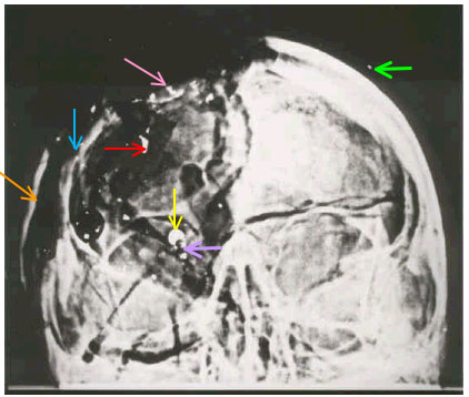
| Figure 1. The AP skull X-ray. Note the 6.5 "metal" object within the upper right orbit (vertical yellow arrow). The elongated fragment (7 x 2 mm), lying above and to the viewer's left of the 6.5 mm object (horizontal red arrow), was authentic and was removed by Humes. The trail of debris (oblique rose arrow), in turn, lies above this, at the very top of the skull. The single, tiny piece of shrapnel in the left scalp is indicated by the horizontal green arrow. Speer's "wing" is identified by the oblique orange arrow (right side of skull). The residual right lateral skull is identified by a vertical blue arrow. Metallic debris (claimed by Speer not to exist) just inferior to the 6.5 mm object, is identified by a horizontal lavender arrow. Some of these (lavender) fragments may have correlates on the lateral X-ray, which would then mark them as authentic metal debris. |
|
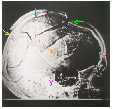
| Figure 2. The right lateral skull X-ray. Note the faintly visible, tiny metal fragment (OTF, i.e., outer table fragment) at the far rear (oblique yellow arrow), just inferior to the discontinuity (fracture). This fragment correlates with (part of) the 6.5 mm object seen on the AP X-ray. The 7x2 mm fragment, removed by Humes, is at the very front (horizontal red arrow). The single, tiny piece of shrapnel high in the left scalp is indicated by the horizontal green arrow. The external auditory canal (large dark dot) is identified by a vertical pink arrow. The oblique orange arrow in the center identifies Speer's "wing." The vertical blue arrow (near the top) identifies Speer's "large disintegrated fragment" (LDF). Two tiny metallic-like fragments (invisible here – lavender arrow) can be seen on the X-rays at NARA, near the inferior pole of OTF. None, however, lie inside of OTF. Furthermore, some of them may have correlates on the AP X-ray, near the 6.5 mm object, which would then mark them as authentic, tiny metallic debris. |
This is a clear mistake all right, but one by Speer, not by Humes. The pathologists were hardly the only ones to view the X-rays that night. While in the morgue, these images were on public display, where many attendees saw them and commented on them. But no one ever described the 6.5 mm object that night. And that was the whole point of the exercise – surely someone would have pointed it out. Even my son (at age 6) and daughter (at age 4) both easily identified it as the dominant feature of the AP X-ray (neither one was then board certified). When I asked Ebersole about it, he abruptly – and forever – stopped talking about the autopsy (listen to my taped interview at NARA). The explanation is simple – it was not there that night. Larry Sturdivan has his own idea: he does not regard this thing as metal (I agree). Instead, he describes it as an artifact (it is), although he seems a bit lost about how that happened (he is not alone). Furthermore, even if Sturdivan were right about this – and it was present that night – how in the world did everyone overlook it? Sturdivan does not comment on this. Even the ARRB experts (see my note 3 above for a reference) all emphasized the gross inconsistency (in optical density) of this thing as viewed on the AP X-ray vs. its partner image on the lateral X-ray. Furthermore, they all agreed on how to correlate its image on the AP with its image on the lateral X-rays, i.e., the 3D coordinates of the 6.5 mm object correlated with the fragment at the rear of the skull. (In my Figure 2, I have labeled this latter object as OTF – for "outer table fragment" – a phrase that derives from the Clark Panel.)
Such a gross inconsistency in optical density had never before occurred in forensic radiology. But the ultimate proof of this gross violation of basic radiology principles lies in the optical density (OD) data. Subjective opinions of the X-rays come cheap, but the OD measurements thoroughly validate these conclusions of gross inconsistency – and they do so in a quantitative (and potentially reproducible) fashion. These results were published in Assassination Science (James Fetzer 1998, pp. 120-137). Regrettably, except for incorrectly using one graph below, Speer does not address these OD data, nor does he offer even an opinion on why they might not be reliable. These data show that the 6.5 mm object (as seen on the AP X-ray) must be longer (from front to back) than all of JFK's dental amalgams stacked side by side – which is an obvious paradox. Aside from photographic superposition (in the darkroom) of this 6.5 mm object onto the AP X-ray, no one has even begun to explain that curious fact. Speer has now joined a large congregation of onlookers who have remained literally dumbstruck by the paradox of this 6.5 mm object. As just one example, John Fitzpatrick, the ARRB's forensic radiologist, who reviewed the 6.5 mm object on two different days, "...continued to be disturbed and puzzled by the fact that the large radio-opaque object in the AP skull X-ray could not be located on the lateral skull X-rays." See my Appendix 1 here for a summary of his findings. Even David Davis of the HSCA (p. 10) had trouble with these X-rays; he said, "It is impossible to work this out entirely."
Probably not. Speer identifies the "next largest fragment" as lying on the main trail (see his figure on his p. 1). See my Figures 1 and 2, where I have labeled this fragment as LDF (for "large disintegrated fragment"). In my opinion, Sibert and O'Neill's description is too vague to interpret with certainty, but the outer table fragment (OTF) would, in common parlance at least, be described as lying at the rear, whereas LDF would be described as near the top of the skull (or near the crown, as Speer says). Fortunately, we don't really need to rely too much on Sibert and O'Neill in this matter, so let's move on.
Yes, most likely he was, perhaps by even more than one. Howard Donahue (whose home I once visited) lists the evidence for these events (Mortal Error 1992, Bonar Menninger). OTF is a good candidate for this. Another is a small fragment near the top of the scalp – on the left side (see Figures 1 and 2). This latter one is visible on both the AP and lateral skull X-rays, even in poor quality prints, and it does lie way off the main trail of debris. Its appearance on the extant X-rays (as viewed at NARA) is totally consistent on the two views and also strongly suggests a metallic fragment. Furthermore, there are even other candidates for ricochet fragments (they are well off the main trail of debris), which I have observed at NARA. Also see my comments under Figures 1 and 2 about very tiny metal fragments near OTF (on the lateral X-ray) and also near the 6.5 mm object (on the AP X-ray). (For data on ricochet angles, see "FBI: Bouncing Bullets." FBI Law Enforcement Bulletin. S. 2-6 u. 20-23. Washington, Sept/Oct 1969. A more recent article is by L. C. Haag, "Bullet ricochet: an empirical study and a device for measuring ricochet angle." AFTE Journal 7 (3): 44-51, December 1975.) Whether such bullets must have struck James Chaney (as Speer insists, albeit without any analysis) would depend critically on the origin of the shot (Speer only mentions the sniper's nest) as well as its timing. Chaney was a motorcycle man located to JFK's rear; his Wikipedia entry describes him as the closest witness to the assassination – except for the limousine occupants. However, Speer is correct to cite Vincent DiMaio and to conclude that ricochet bullets do not break into narrow cross-sections or slices (even though Speer promptly introduces his own slice). He is also correct to confirm that the nose and tail of the bullet (which supposedly deposited the 6.5 mm object) were both reportedly found in the limousine. Unfortunately, since he has just quoted DiMaio, Speer sows confusion when he apparently states the opposite:
Even more puzzling, he seems to reverse himself once more on the next page (p. 4): "...it makes little sense to believe that the middle of a bullet...would get sliced off upon entrance to the skull...". I think that what Speer means is that a slice can arise after entering the skull, but not at the point of entry. But he does insist that the 6.5 mm object represents an authentic piece of metal, one that came from the "middle of the bullet." That is, of course, an extraordinary denouement – unsupported by any forensic data, and surely not approved by DiMaio. Here is what the HSCA's ballistics expert (Larry Sturdivan) thinks of this proposal:
To make matters even worse, since Speer claims that the JFK X-rays are authentic, he must also believe that this 6.5 mm object was indeed present on the AP X-ray that night – but that no one noticed it. Speer totally evades this profound conundrum, as if he were blissfully unaware of it.
Speer also quotes from DiMaio (Gunshot Wounds: Practical Aspects 1985, p. 90), who reports no ricochet from a 6.5 mm full metal-jacketed bullet for impact angles of 20º and 30º. The following data (from the same table), however, are omitted by Speer. For this same bullet, a ricochet angle of 1.6º results from an impact angle of 10º. In addition, for impact angles of 30º, various other bullets yield ricochet angles of 1.19º – 2.48º. DiMaio also adds that partial metal-jacketed bullets usually break up on impact and then pepper the body with fragments from the jacket or from the core. He notes that these projectiles typically lodge in or just beneath the skin (that reminds me of JFK's back wound). The multiple, tiny metallic fragments I saw in the skull X-rays (and the shallow projectile that caused the back wound, too) might thus be explained via such ricochet, but Speer carefully avoids following DiMaio down that path. Several pages later (p. 12), Speer notes that the nose of the bullet (CE-567) was covered with skin [for laboratory analyses of evidence released by the National Archives and Records Administration (NARA) click here], so the question naturally arises: Was this the projectile that caused JFK's back wound? The problem, of course, is that this nose fragment was officially discovered in the front seat of the limousine so, unless some mix-up later occurred, that explanation won't work.
Since Speer regards the brain photos as truly JFK's, he needs to square this comment with the nearly intact right brain seen in the autopsy photos. Unfortunately, he totally evades this issue. In fact, the OD data demonstrate that a good deal of the right brain was actually missing (which is consistent with the Parkland observations). Ultimately, however, this question cannot be answered – because authentic photographs of the brain no longer exist (Inside the Assassination Records Review Board 2009, Douglas Horne, Chapter 10).
Perhaps it does. Speer cites a peer reviewed article (Radiology 240; No. 2, pp. 522-528, August 2006), in which this occurred in 8 of 10 cases, but he omits the following details. This study included 78 wound tracks in 13 cases, i.e., about six per person (which is clearly different from JFK). All subjects were injured by high-velocity 7.62 mm bullets from an AK-47 (probably also different from JFK). The authors admit that decompositional changes (especially in the brain) could have affected their interpretation. In particular, a distinct linear track within the brain could not be identified in any case. In addition, they emphasize that their small sample size limited their conclusions and they reported that their results would still need to be confirmed in a larger study. I would add that Doug DeSalles and I do not recall a similar outcome (of such brain settling) in any of the nineteen (19, not 9) cases we reviewed (of fatal gunshot wounds to the skull). Also, as best I can now recall, our cases typically had suffered only a single head shot. If such a CT scan study had been available for JFK, many of today's mysteries about his skull trauma would have vanished; in particular, a 3D reconstruction of a skull (in this Radiology article) shows a remarkably detailed image of the comminuted skull fragments and skull fractures.
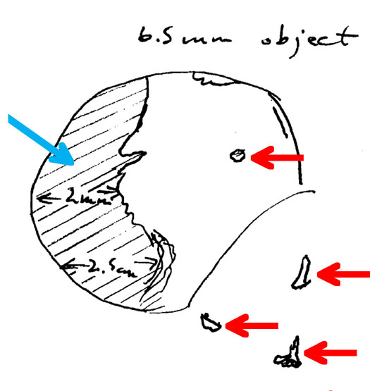
But it did leave small fragments! See my slide 33 from the Dallas lecture or my Figure 3 here. My sketch shows tiny metallic debris lying immediately inferior to the 6.5 mm object and at least one piece (paradoxically) inside the 6.5 mm object! (There may be more inside.) These (exterior) pieces can actually be seen in my Figure 1 (horizontal lavender arrow). These observations were made before my Lasik surgery, when I was extremely myopic (-9 diopters) and I could see such small objects in amazing detail without eyeglasses. That these things are metal is strongly suggested by the lateral X-ray, where two tiny fragments lie near the inferior pole of OTF (but outside of it). These two may well have correlating images on the AP X-ray; such a correlation would virtually guarantee their authenticity as metallic debris (presumably from a ricochet). If OTF is authentic, no other fragments should be seen superimposed over the inside of it; in fact, none are (which is different from the 6.5 mm object seen on the AP X-ray).
|
| Figure 3. This is a magnified view of the 6.5 mm object, as I sketched it in my NARA notebook; I was still very myopic at that time so I could see nearby objects in remarkable detail. Notice the three fragments immediately outside of it and at least one inside its borders (all identified by red arrows). In addition, note the original, authentic fragment (cross-hatched – oblique blue arrow), which was probably described by the FBI. This one correlates with OTF (outer table fragment) seen on the right lateral skull X-ray; both the size and location (of the cross-hatched fragment) match to OTF. With my naked and myopic eyes I could actually see this cross-hatched, authentic fragment as an optical superposition. Speer fails to locate OTF anywhere on the AP X-ray, but he is apparently unaware of this gaffe. (This figure is similar to slide 33 in my Dallas lecture.) |
Here is Speer's actual comment, which is clearly wrong (about no small fragments located near the 6.5 mm object):
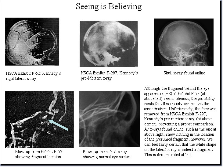
So Speer seems to say that the 6.5 mm object arose near the exit site, after which it presumably (in his scenario) traveled to its final resting site, where his "slice" is now seen in my Figure 4. (A bit more clarity from Speer would help here.) More importantly, however, he offers no evidence whatsoever from forensics that such an event is even possible.
|
| Figure 4. Seeing is Believing. This figure is copied from Speer's p. 21. The oblique blue arrow (Speer's arrow, not mine) identifies his "slice," which (he claims) correlates with the 6.5 mm object on the AP X-ray. Of course, by reaching this conclusion, Speer has only created another paradox – he leaves OTF (outer table fragment) without a correlating image on the AP X-ray, but he seems unconscious of this. |
It is true that Robertson and Riley disagree with me. (I don't know who Burnett is, but Speer cites him as a radiology colleague of Randy Robertson.) However, neither of them has attempted to explain any of the paradoxical OD data. For that matter, no professional has even tried to explain the obvious paradox of the 6.5 mm object as viewed on the AP vs. its corresponding image on the lateral – the ARRB experts are excellent examples of this (failure to explain). In the history of forensic radiology, this is a unique event. It is true that there is no published list of radiologists who support my view. However, my best friend (a superb diagnostic radiologist), who played a critical role in illuminating how X-rays were copied in the 1960s, is a strong supporter of my views. Dr. Siple (whom I met), a friend of Harry Livingstone (see Siple's comments in Harry's books), had long suspected that the X-rays were composites (Assassination Science 1998, p. 156), which matches my own view. My Dallas lecture also cites Arthur G. Haus, the chief medical physicist at Kodak (whom I have met), as not offering any critique of my original OD paper; on the contrary, he found it very interesting. (Of course, as a then-Kodak employee, he could not make comments that might affect his own company.) But the chief problem, as Speer himself notes (based on his own online efforts), is a lack of interest by diagnostic radiologists; by his own report, he apparently got feedback from only one, and that discussion did not relate to the OD paradoxes. Finally, I did eventually receive a letter from the ARRB's forensic radiologist, John Fitzpatrick, in which he made no substantive comments and firmly declined to discuss the JFK X-rays (Appendix 2). So the matter rests.
This is a common allegation, but it is misleading. Speer also buys into this myth – he even castigates Custer and Reed for screwing up so badly (even though, paradoxically, he later prefers their opinions – on other matters – over that of true experts). Think about this: no one claims that the other X-rays (of JFK's extra-cranial sites) were likewise overexposed. They weren't. So why would the skull X-rays alone be overexposed? Actually, they are not. It is common practice for X-rays to contain optical densities in the range of 0.5 to 2.0, so that the human eye can make rather easy distinctions among different densities. In fact, except for the Dark Area, most of the ODs on the JFK skull X-rays do lie within that typical range, as I have verified via hundreds of measurements. Even the densest bone (the petrous) falls within that range. Back when I first viewed them, the skull X-rays did not strike me as overexposed. Furthermore, such a response has not characterized other viewing experts either, e.g., none of the ARRB experts said that. One exception to this is Russell Morgan, who called them "severely overexposed" (p. 17). [Morgan was the forensic radiologist on the Clark Panel (named for US Attorney General Ramsey Clark), which published its report (of no conspiracy) on January 16, 1969, just days before the Garrison trial began. This panel was led by Russell Fisher, a forensic pathologist. Curiously (some would say suspiciously) both Russells had offices at Johns Hopkins University; in addition, the report had been long delayed, perhaps to counteract the Garrison trial. The Clark report can be found here.] However, with less exposure, the White Patch would be even whiter – and its OD would fall below the normal range for viewing X-rays! Interestingly, Morgan chose to ignore this absurdity. The HSCA, of course, enhanced the X-rays, but I suspect that was mostly to obtain useful prints for publication. (Printing changes the contrast.)
Speer claims that I failed to discuss issues of contrast in the JFK X-rays, thereby imputing this supposed failure to my specialty as a radiation oncologist. By contrast, Speer favorably quotes another radiation oncologist (John Ebersole) but then generously overlooks his specialty (which was the same as mine). More to the point, though, Speer ignores my history as a physicist, which is actually far more germane to this matter than is my specialty. (Just ask a random diagnostic radiologist some detailed OD questions, especially about characteristic curves, if you seek proof of this.) I had, in fact, addressed these issues in some detail in a rather long, but unpublished manuscript (privately circulated in 1994). Many pages were devoted to technical issues regarding OD, including characteristic curves of X-ray films. Although Speer is probably ignorant of this history, he failed even to be curious about it, and instead falsely accused me of being superficial.
Now one final point should seal the deal. I measured the ODs in the background of these X-rays, where only air surrounds the body. These background ODs provide a very useful check on the relative exposure of one X-ray film compared to another. The ODs quoted here are based on several measurements (up to ten) for each X-ray, but the range of ODs on each one was narrow. Here are the mean ODs: AP skull = 3.99; right lateral = 4.01; left lateral = 4.18; abdomen = 3.75; pelvis = 3.73. This represents only a modest range of exposures among the different anatomic sites. The one outlier is the chest, with a mean background OD of 3.42. This implies a lower exposure, but since lung tissue does not need as much exposure, that would be expected. In fact, to use the same exposure for the chest as for the pelvis or abdomen would lead to an overexposure. In short, all of these numbers fit together very well and are not at all surprising. Further support for this conclusion comes from John B. Cahoon (Formulating X-ray Techniques 1966, pp. 167-168). Suggested exposures for the abdomen, pelvis, and skull are almost identical: for the same current (100 milliampere-seconds), they differ only modestly in voltage (respectively 62, 64, and 70 kV). By contrast, the suggested PA chest exposure is only 10 milliampere-seconds (at 62 kV), a much lower exposure. These exposures are completely compatible with the background ODs on the JFK X-rays. Therefore, to claim that the skull X-rays were incorrectly exposed (and also to accept that the extra-cranial X-rays were correctly exposed – which they were) makes no sense. This discussion should just be put to bed – and Morgan was wrong to say that overexposure had occurred. The OD data convincingly close this case.
No – definitely not. This is an eccentric charge by Speer, and it reflects badly on his approach to this subject. At NARA, I used only the extant X-ray films, not prints and not enhanced X-rays. In fact, while at NARA I never even viewed prints of X-rays or any enhanced X-rays. It is true, though, that the published prints of the JFK skull X-rays have been enhanced, but that is because the prints of the unenhanced X-rays do not accurately portray the extant X-rays. In print format, the enhanced X-rays are closer in image content to the extant X-rays. Since Speer had been exchanging e-mails with Fetzer (he quotes Fetzer), he could easily have asked Fetzer (about whether I had used the extant X-rays), but he forgot to ask. Of course, Steve Tilley (and Gary Aguilar, too) can also verify exactly what I used. Speer concludes with this statement:
How Speer reaches this remarkable conclusion, without once addressing any actual OD data, he does not explain. Even if Speer were ultimately to prevail here, such opinions, reached without serious underpinnings, cannot become candidates for serious conversation. He could, at the very least, offer an opinion on why the ODs of the White Patch are similar to those of the petrous bone (in the right lateral X-ray) – after all, three layers of bone will not explain this. Another troubling paradox for Speer is that the White Patch and the petrous bone are not nearly so similar to one another (in OD) on the left lateral skull X-ray. Of course, this might well have occurred if the double exposure – of the fake White Patch – had been somewhat different on the two lateral X-rays.
This is the easiest question of all; just think – if forgery had occurred, then that is precisely the expected (and almost certain) outcome! On the contrary, with honest X-rays no such persisting confusion should ever have arisen. Notice, in particular, how the 6.5 mm object greatly troubled John Fitzpatrick (the ARRB's forensic radiologist) – so concerned was he that he even returned to it for a second day, yet he never could explain it. Speer does not address issues of authenticity in any detail, which – in view of Fitzpatrick's failure to solve the puzzle – should scarcely surprise us. Speer then cites his reluctance to
Speer then quotes at length from Horne (who was citing me): in short, I stated that the HSCA site shows no entry (as confirmed by the OD data, a basis that Speer ignores), but Speer claims that this conclusion is evidence of my belief in an exit high on the rear of the head. He finishes by suggesting that the HSCA entry site may be real, but merely be located somewhere else! (No evidence is offered for this.) Here is my response to this semantic bog.
Via detailed OD measurements, I was not able to locate a hole at the rear of the skull anywhere near the HSCA entry site. And where the main trail of debris projects to the rear of the skull, the AP X-rays suggest no skull bone, so it is natural to assume that some debris did exit there. However, in the absence of skull bone, one surely cannot expect to see a "hole" in that vicinity. That some debris did, in fact, exit to the rear, where it struck the follow-up limousine and at least one motorcyclist, seems quite certain. The other option for such an exit, of course, is the hole in the right occiput, as reported at Parkland. This is, of course, much lower than the main trail of debris. As expected, Speer does not mention this latter site as a possible exit.
No – definitely not. The dictionary definition of "distortion" is a "change in shape." What Speer actually means is magnification, which is quite another matter. Magnification alone does not change the shape of an object. Although magnification does affect these X-rays, that effect is easily manageable.
Surely not. The so-called "slice" that Speer identifies on the lateral X-ray (my Figure 4) is the ultimate "boner" (Speer himself introduced this pun – see p. 18). No expert has ever identified that site as a piece of metal. Even Speer, if he had viewed the extant X-rays, would not have made such a blooper. The discussion that follows from his misidentification should just be ignored – totally. The reader should simply ask himself a simple question: Who is more likely to be correct – an amateur who has viewed only prints or zillions of experts, who have seen the X-rays? It is true that phrases (some by Humes, but others have contributed, too – see pp. 24-26) have imprecisely located the 7x2 mm fragment (Speer's club), but the bottom line is simple: despite the semantic fog, there is really only one large metallic fragment under discussion – and it's not the "slice" cited by Speer. His "slice" is just a bone spicule, certainly not metal. It has nothing to do with the case, except that it might have resulted from trauma. The only authentic large metal fragment involved in the autopsy is the 7x2 mm one (identified in my Figures 1 and 2), which Humes removed. Speer might also want to read again his own quotes from Humes (p. 25), about the 6.5 mm object: "I can't be sure I see it in the lateral at all, do you?" And this one too: "I don't remember retrieving anything of that size. "
Yes – definitely! If you disagree, then try this question: Given the metal fragment at the rear (OTF) of the right lateral X-ray, where is its correlate on the AP? I have never found anyone who can answer this question – unless it lies (paradoxically) inside the 6.5 mm object. And that is precisely what my myopic eyes saw at NARA – an optical superposition of the faked 6.5 mm object over the underlying authentic fragment at the rear of the skull (OTF).
No, he has not. John Hunt has summarized sample-size requirements (private communication):
Hunt discovered that only 2 mg was actually taken for spectroscopy. This is only a tiny fraction of the original mass (106.92 mg) of the larger fragment.
Speer claims that I insist the 6.5 mm object is not visible on the back of the head. This is scarcely an accurate portrayal of my work. On the contrary, I have repeatedly stated that the location of OTF (on the lateral X-ray) correlates extremely well with the 6.5 mm object on the AP X-ray. So do virtually all experts who have viewed these films. The real issue is slightly, but seriously, different: Are the ODs of this thing consistent from one view to another? That answer is clearly, "No," as even the ARRB experts readily emphasized. But Speer is relentless – he then also takes Horne to task for misrepresenting the situation. Somehow, though, Speer has still missed the point – it's all about the inconsistent ODs, not the 3D coordinates (which do match). But then, strangely enough, Speer notes my "...near religious belief the fragment in the AP X-ray [the 6.5 mm object] has been added atop a much-smaller pre-existing fragment...". So it seems (at least semantically) that he can state my proposal, despite his earlier misrepresentations. Unfortunately, as before, Speer does not even begin to address the actual OD data that support my conclusion (of superposition). That the OD data (presumably hard science) provide the basis for my "religious" belief, according to Speer, is especially ironic. Oddly enough, if he had known of my remarkable religious pilgrimage, he might even have winked at me while making such a statement. But let's put the chief question directly to Speer: If OTF (on the lateral X-ray) does not match (in 3D) to the 6.5 mm object (on the AP X-ray), then where we do see the correlate of OTF on the AP? If I could choose one question for Speer to address, this is it. To date, no one has dared to answer this question. Speer, of course, has chosen to match his "slice" (on the lateral X-ray) with the 6.5 mm object (seen on the AP). But that leaves OTF without a partner on the AP X-ray, which is surely a unique event in the history of radiology.
Yes, of course it was. This size was cited by both the HSCA and the Clark Panel. That size is merely based on a physical measurement (no magnification correction) on the AP X-ray, which is a trivial matter. Since this thing correlates with the metal at the rear of the right lateral skull X-ray (OTF), then magnification should be not an issue (because OTF lay adjacent to the film). Of course, if my proposal of photographic superposition is accepted, then magnification is quite irrelevant. Speer cites my OD graphs (and displays one of them), from which he extracts a width of 7.4 mm. His measurement technique, however, is highly unorthodox. Most scientists would measure from the halfway point (between minimum and maximum ODs) at either end of the curve: that yields a width of 6.5 mm, which agrees with measurement directly on the extant X-ray film. This is hardly news – I had made that determination from the graph immediately after recording the data.
Chapter 18b: More Fun with X-rays
No, it does not – nor could it even do so in principle. First, these are two distinctly different areas, as should be obvious from the right lateral X-ray – the White Patch is much more posterior than the overlap area. See my image of the White Patch in Assassination Science 1998, p. 160, or slide 5 in my Dallas lecture, or my Figure 5 just below. Speer does not display my image, but he should have. For comparison, Speer displays his "wing" on his p. 7; that image is copied here in Figure 6.
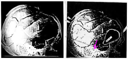
|
| Figure 5. Right lateral X-ray showing the White Patch and the Dark (Frontal) Area. For the image on the right, I have circled (black dotted line) the White Patch, but it is readily apparent, even to the naked eye, on the left image. Also note the absurdly identical whiteness (on the left image) in the petrous bone and in the White Patch. On the right image, the petrous bone (which surrounds the external auditory canal – pink arrow) is faintly circled, while the Dark Area is circled in white. The external auditory canal locates the approximate center of the external ear (see my Figure 2). |
|
| Figure 6. Where is the "Wing"? This image is copied from Speer's p. 4. He locates the "wing" between the two red lines. Presumably (although Speer does not state this clearly) the "wing" is identified by red shading. Notice that the "wing" lies directly superior to the external auditory canal (the latter is identified in my Figure 2), which is the approximate center of the external ear. |
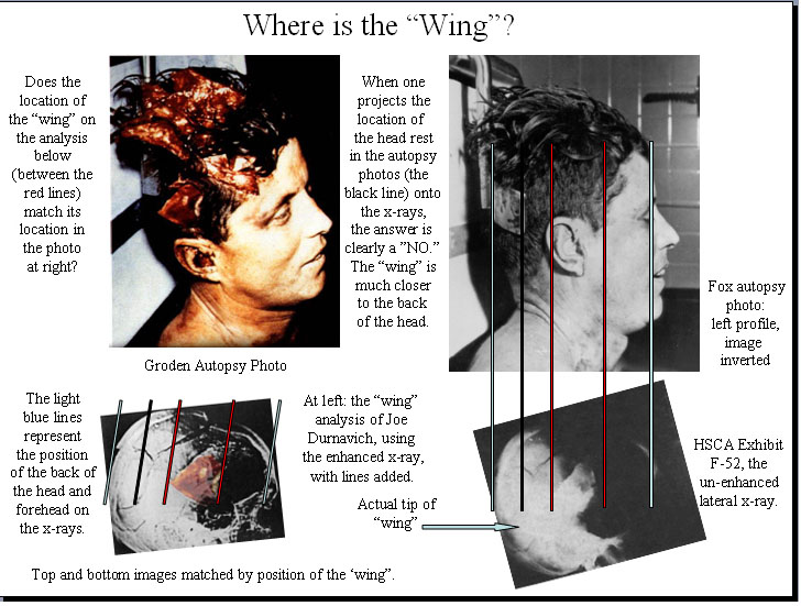
In his image (my Figure 6 here), Speer locates the "Actual tip of ‘wing'," presumably meaning its most posterior tip (although his syntax is fuzzy). Even if that unreasonably far posterior location is accepted, it is still far too anterior to match the posterior border of the White Patch. The location of the White Patch, especially its posterior border, has repeatedly been confirmed by the OD data – it does not depend on the human eye (although it does match what the eye sees); in fact, the whitest area lies immediately anterior to the inner table of the occipital skull, well posterior to anyone's location for the "wing." Furthermore, it is visibly obvious (see Speer's images) that the "wing" lies superior to the external ear and cannot extend far posteriorly. In my Figure 2, I have identified the external auditory canal, which Speer ignores; that structural feature clearly locates the external ear – without any ambiguity. Speer also ignores the evidence of the AP X-ray (my Figure 1). Notice there how the wing lies far out in space, quite detached from the skull. On the other hand, if the wing had extended far posteriorly (as Speer wants to believe), then some part of it would be seen much more medially in the AP X-ray, but it is not there. This argument is so powerful that little else need be said. But there is more.
Second, the ODs of these two areas are quite different: on the right lateral X-ray, the mean OD of the white patch (0.625 ±.055) is almost the same as the petrous bone (0.55), whereas a typical OD (1.33) for the overlap site is noticeably higher (than the White Patch), and it does not appear nearly so white to the eye. That visible difference is dramatically obvious in Figure 5 (especially on the right sided image). Speer claims that the White Patch was caused by three overlapping layers of bone. Despite his unrelenting caricature to the contrary, I have always accepted three layers of bone at the overlap site, although I have never emphasized this because no one (before Speer) had offered such a novel explanation for the White Patch. Incidentally, the three layers of overlapping bone should be obvious to anyone after viewing the AP X-ray (an image that Speer overlooks). He also argues that, because the ARRB experts (p. 10 and also Chapter 19b, pp. 26-27) noticed such bone overlap, they therefore support his conclusion that the overlap explains the White Patch. But that is simply absurd. We all (even me) understand that bone overlap (of three layers) is present. On the contrary, the question is this: Does the overlap explain the White Patch?
Third, the White Patch is so dense that whatever physical object it represents must appear somewhere on the AP X-ray film. I made this argument from the very beginning, even at our first press conference in New York City (1993). That transcript is reproduced in Assassination Science 1998 (p. 155) and warrants a quote here:
No one has even tried to explain this paradox. Even worse, Speer seems oblivious to it.
Let's next focus on the OD issues for overlapping bone, a quantitative exercise that Speer totally neglects. For these JFK skull X-rays, here are the pertinent OD changes (∆ODs) across various layers of bone: one layer = 0.45; two layers = 0.90; three layers = 1.35. The difference for one layer is easily measured at fracture lines; amazingly enough, Speer believes that I ignore these fracture lines (p. 9). If an extra bone layer truly explained the White Patch, then sites just outside the White Patch should yield ODs that are higher by about 0.45 (one layer). But that is not the case – on the contrary, the ODs suggest a difference of more than just one layer of bone. Of special interest is the OD over the occiput, at the very back of the skull (very close to the White Patch), where the bone is viewed tangentially: the data there suggest a ∆OD (compared to the White Patch) of not just more than one layer, but actually about two bone layers (i.e., it is much less white). In other words, the White Patch is truly an anomaly (much too white and with ODs that are far too low). It cannot possibly arise simply from overlapping bone. On the other hand, of course, a deliberate superposition of this area in the dark room could easily explain this paradox. That the ODs of the White Patch and the petrous bone are not nearly so identical (to one another) on the left lateral X-ray should also raise some doubt that not all is well in OD land.
Now recall that three layers of bone yield a ∆OD of 1.35. Since the measured OD (cited above) in the overlap area is already 1.33, the OD without the three layers of bone would be 1.35 + 1.33 = 2.68. The ODs in the maxillary sinuses (mostly air) are 2.89, so this value of 2.68 clearly suggests substantial missing brain in the overlap area. But the site in question (medial to the overlapping bone on the lateral X-ray) lies near the middle of the brain, where the autopsy photographs show no missing brain tissue! Also recall that the pathologists described the brain laceration as only 4.5 cm deep, which would lie just above the "wing." (This level is demonstrated on the right lateral skull X-ray in the DiEugenio reference at the end of my Appendix 1 – see Figure 5A in that article.) Since Speer believes the autopsy photographs of the brain are JFK's, this missing brain poses yet another paradox for him, which, of course, he does not address. The bottom line is that, given his state of knowledge, Speer has offered a zealous, honest and original proposal, but edicts are not evidence and proclamations are not proof. A thorough analysis of all of the data is always required. Moreover, he had seemed to agree with me (p. 5) that large dark areas (not merely fracture lines) represent missing brain, because the brain typically contributes much more to the overall OD than does bone, but in this specific discussion he has forgotten that lesson (or perhaps he changed his mind without telling us).
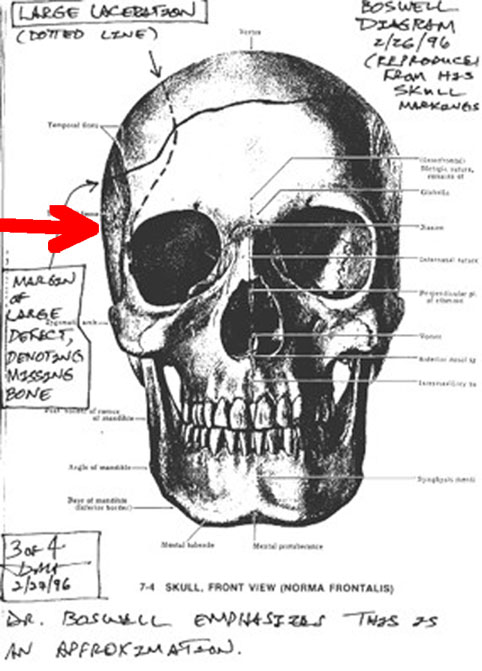
Speer also claims that the Dark Area contains only one layer of bone. Even a brief look at the AP X-ray, though, shows that this is most likely wrong. In my Figure 1, residual bone along the right lateral skull is indeed present (vertical blue arrow), and so is the symmetric bone on the left side; therefore two layers are present. Furthermore, Boswell's autopsy diagram (cited by Speer, or see slide 23 in my Dallas lecture) clearly shows bone present on both sides of the skull in this region. Boswell's skull drawings for the ARRB also confirm this (see my Figures 7 and 8 here). By simple logic therefore, the large Dark Area did not result from having only one layer of bone; it actually has two layers. On the contrary, the darkness must represent a large volume of missing brain. Moreover, Speer's quoted Radiology article (if he accepts its conclusions) offers compelling evidence for just such missing brain at this anterior site (in those cases), but he seems to have forgotten what he read there.
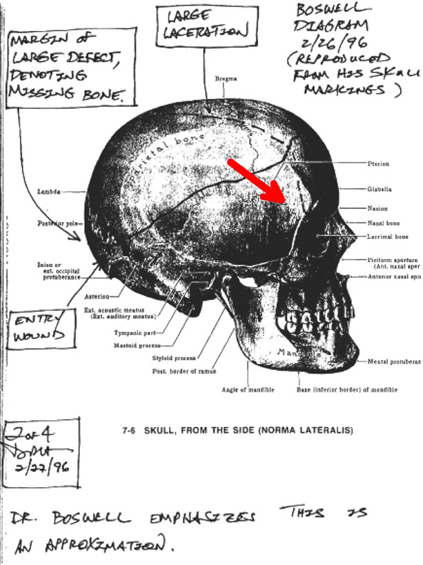 |
| Figure 7. Boswell's drawing on a skull – lateral view. Doug Horne copied (onto a piece of paper) Boswell's drawing on a 3D skull for the ARRB. Notice, in particular here, how much bone is present on the right lateral skull, in the region of the Dark (Frontal) Area (arrow). The latter phrase is my description of this dark region as seen on the lateral skull X-rays (both right and left). |
Chapter 19b: Stuck in the Middle with You
|
| Figure 8. Boswell's drawing on a skull – AP view. This is the AP view of the same skull drawing by Boswell. Notice the presence of bone on both sides of the skull, where the Dark Area (arrow) would appear on the lateral skull X-rays. |
Yes, we agree! But if it was, why is Speer so certain that the medical evidence is so pristine?
20. What does the Harper fragment tell us? (pp. 21-25)
I have already offered my apology for confusing the audience with the site of the metallic debris on the Harper bone. Even though it is decisive, Speer does not cite the Harper X-ray at all, even though I did show the close-up view in Dallas. Using the Harper photographs, I had placed this (presumably) lead debris at one corner of the fragment. See Speer's reproduction of the "Mystery Photo F8" (p. 21), or see slide 22 in my Dallas lecture. In retrospect, I don't actually know which site the Dallas pathologists had picked, nor have I ever met anyone who knew. I only knew that they had picked some site. Just based on the photograph, though, the site I originally selected had looked suspicious to me, and, without the X-ray, I might still pick it today. The reader may wish to try this exercise himself, or even try it on friends. Quite amusingly, Speer (p. 24) also places the lead debris where I originally did! The Harper X-ray, however, shows the lead debris at the opposite pole of the fragment. See my Figures 9 and 10 here; the X-ray images are courtesy of John Hunt.
|
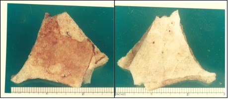
| Figure 9. Harper fragment photos from the Dallas pathologists. The outer surface is on the left: note the faint lead smudge (red arrow) at the upper left, at the very edge. The inner surface is on the right. No evidence for metal of any kind is seen on this inside surface. |
|
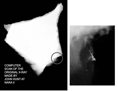
| Figure 10. X-ray of the Harper fragment. Note the metallic debris, circled on the left, and shown enlarged on the right. This is the same site as the lead-like smudge that is identified on the photograph in my Figure 9 – just rotate either photo by 180º for easier comparison. John Hunt is acknowledged (and thanked) as the source for this X-ray, which he discovered at NARA. |
Whether any metal is present at my originally selected site may not even be finally answered by the X-ray, but, in principle, it might have been decided by other physical and/or chemical tests performed on the actual bone (which is now long gone). For the present, therefore, we are stuck with the X-ray evidence. In his essay, Speer displays my placement of this fragment (p. 23) in the "Overhead View of Human Skull" from my Dallas lecture (slide 20). Notice where I have labeled "Metal debris – confirmed." This is the metallic site identified in the Harper X-ray (Figure 10). On the exterior surface in the photographs (Figure 9) there is a suggestion of lead at the same site as the X-ray. If that evidence is accepted, then Angel's (and Riley's) placement of the Harper fragment (my Figure 11 here) does not make any sense. I had deliberately placed the Harper fragment (slide 20 of my Dallas lecture) deliberately too far to the right (for Angel's placement), just because I did not want to obscure the sagittal suture. On the contrary, to correctly mimic Angel's conclusion, the "Suture line, according to Lawrence Angel" should exactly overlap the sagittal suture (as it does in Angel's sketch in my Figure 11 here). Of course, I do not accept Angel's interpretation. Instead, the Harper fragment most likely came from the high occipital area, as I have argued elsewhere.
|
| Figure 11. Angel's placement of the Harper fragment (in blue). The delta fragment here (in red) lies anterior to the coronal suture (probably in its correct location). Note the suture line on the Harper fragment, a structure that Angel did accept. I borrowed this colored sketch from John Hunt; the uncolored version was published by the HSCA. The red arrow points at the metal debris (on the outside of the Harper fragment), based on the Harper X-ray. |
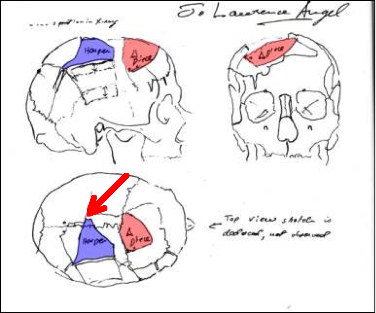
According to Angel, the sagittal (i.e., midline, top of the head) suture is visible on the Harper fragment. That suture line helped Angel to locate the Harper fragment near the skull vertex, as shown in my Figure 11. However, based on the Harper X-ray, the lead site then lies just to the left of the skull vertex – and the lead is on the outside of the skull! That is truly bizarre. No one has ever proposed that a bullet entered at this site, yet that is precisely where Angel's (and Riley's) placement of the Harper fragment has led them. There is even more evidence (in a forthcoming essay) that my placement of the Harper fragment (mostly from the upper occipital area – see my essay in Murder in Dealey Plaza) is correct, after all. However the bottom line here is this: if one accepts the Harper X-ray evidence, then the Angel location – with lead lying to the left of midline on the outside – cannot possibly be correct. Angel, however, can be forgiven. He was told, as a fait accompli, that the occipital bone was intact, so he had little choice about where to put this bone. Also, even more importantly, he knew nothing about the Harper X-ray, but now everything has changed.
On Closed Minds
Speer adamantly claims that most characters (on both sides of these JFK debates) have totally closed minds, which they won't change for anything (p. 27), a category into which he presumably dumps me. He had earlier (p. 9) also cited The Structure of Scientific Revolutions (Kuhn 1962) to the same effect. But I plead not guilty to his charge – Speer should think hard about the following facts. First, at a rather early stage in my OD work, after I had (wrongly) decided that the OD data were inconsistent with composite X-rays (in a widely, but privately, circulated paper, titled "2 + 2 = 4"), I had followed the data where (I thought) they led and stated that the X-rays must be authentic. Speer apparently does not know this history. The correction of my mistake came from Arthur G. Haus and colleagues at Kodak, who advised me about image crossovers (from one side of the film to the other) in these 1960s X-rays, a technical problem that was later solved. (Initially, I had only known about modern X-ray films, where the image cannot effectively cross over from one side to the other.) The presence of such crossover in these JFK X-rays, though, re-opened the door to photographic alteration in the darkroom. Haus later read my paper, which discussed these image crossovers in the JFK X-rays. In view of this, Speer is demonstrably wrong to say that my mind has been forever closed. (Regarding the role of irrationality and bias in human decision making, see two excellent references – Irrationality: The Enemy Within 1991, Stuart Sutherland and Persuasion: Theory and Research 1990, Daniel J. O'Keefe.)
Second, as further evidence for my open-mindedness, Speer should review my rejection of the acoustic evidence (a 72-page essay for the CTKA website). A senior JFK researcher (who does not espouse a JFK conspiracy and who I greatly respect) remarked that I am the only conspiracy believer (so far as he knows) who has clearly disavowed the acoustic evidence. (I do not know where Speer stands – or sits – on the fence atop the grassy knoll.)
Third, another event might also give Speer pause: when Fetzer (my own editor and still a dear friend) overstepped the accepted bounds of public civility, I publicly chastised him, an event that Speer also seems to have missed. That sad event displays a lifelong curse: my primary loyalty is to my ideals, even at the expense of close friends (but I would not wish that handicap on anyone else, not even on Pat). In any event, here is the challenge for Speer: if he can truly show me to be wrong, then I shall recant again (of my JFK beliefs, but probably not of my religious views). On the other hand, if Speer were to recognize his imperfections, he would be welcomed back as warmly as the Prodigal Son.
A Few Final Thoughts
Lest there be any doubt, let me be very clear: I admire Pat's passion. We need more Americans like him. And I really think I would like him if I got to know him. I would be remiss, however, if I did not admonish his readers to probe deeply into the foundations of his arguments before accepting his conclusions. In addition, Pat himself might consider becoming a bit more disciplined before careening into verdicts. I would also encourage him to lay aside his ad hominem attacks. David Hackett Fischer (Historians' Fallacies: Toward a Logic of Historical Thought 1970, p. 293) has critiqued such ad hominem attacks: "But an ad hominem debate is unlike tennis in one respect – it is a match which everybody loses: players, referees, spectators and all. " These attacks do not lead to any new knowledge and they surely won't win Pat many new friends. In this tent (of researchers) we have acres of space for divergent views – but tolerance is always welcome. Finally, and more specifically, the implications of the Harper X-ray need to be integrated into our understanding of JFK's skull trauma. My kudos to John Hunt for this wonderful discovery.
Acknowledgments. I am deeply grateful to Douglas Horne and James DiEugenio for their careful reading and valuable comments. I have already noted the essential contributions of John Hunt. It is a luxury to have accomplices such as these.
Appendix 1. My letter to John Fitzpatrick (with attachment)
November 3, 2009 John J. Fitzpatrick, MD Re: JFK Autopsy Skull X-rays Dear Dr. Fitzpatrick: I recently read a staff summary of your medical presentation to the ARRB. In the attachment here I have listed 12 points of agreement. The only possible point of disagreement is not even certain; it is possible that there are no points of disagreement at all. Nonetheless, the ARRB staff summary quotes you as saying that you disagree with my work. I wonder if I could persuade you to be more specific. In fact, there is a specific purpose: I am scheduled for a talk in several weeks on this very subject. The chief medical physicist at Kodak (my own PhD is in physics) read my original paper (regarding the OD data on the skull X-rays), said he found it very interesting, and offered no specific critiques of it. You will also note that Cyril H. Wecht co-authored an article with me (see footnote on the attached page). You may find my latest presentation on this subject online at http://www.assassinationresearch.com/v2n2/pittsburgh.pdf Also see: Mantik, D. W. (2000), "Paradoxes of the JFK Assassination: The Medical Evidence Decoded," in J. Fetzer, ed., Murder in Dealey Plaza (Chicago, IL: Open Court/Catfeet Press, 2000), pp. 219–297. Sincerely yours, David W. Mantik |
The JFK Skull X-rays [also sent to Fitzpatrick]
Fitzpatrick (JF) vs. Mantik (DM): Points of Agreement
by David W. Mantik
October 30, 2009
- The left brain silhouette can be seen in the AP film.
- The extremely dark area on the upper right in the AP film represents missing brain (replaced by air) in an open wound.
- The orbit of the right eye is fractured and displaced.
- No entry wound is seen on the AP film.
- The 6.5 mm object (on the AP) looks metallic.
- The two burn marks (on the AP film) are unique.
- No entry wound is visible on the lateral films.
- No definite object is seen on the laterals to correspond to the 6.5 mm object on the AP film.
- A small object is seen on one lateral film that was spatially consistent with the 6.5 mm object (on the AP), but it was not of the appropriate optical density.
- The small metallic fragment posterior to the right eye on the lateral does not correspond to the 6.5 mm object (on the AP).
- Most missing skull bone is parietal.
- The direction of the bullet cannot be ascertained from the "snow trail" on the lateral film.
- Most of the frontal bone is present, at least up to the hairline.
- From the three bone fragments, it is impossible to determine the nature and direction of beveling.
- Metallic fragments are seen on the largest of these bone fragments.
- A suture, as well as an adjacent break in bone, is seen on the largest fragment, but the specific suture cannot be identified.
Fitzpatrick & Mantik: A Possible Disagreement
JF concludes that the left frontal brain is present. DM reports this: OD measurements on the lateral, through the maxillary sinuses, were compared to ODs in the dark frontal area. These two different sites show ODs that are very similar, which strongly suggests very little soft tissue in either area, i.e., very little brain on either side. One possible resolution of this apparent disagreement is as follows: if the left frontal brain has been displaced posteriorly, both parties could be correct. In fact, DM agrees that this might well be the case. DM only states that the area where the left frontal brain should lie is empty on the lateral films. However, OD measurements on the left side of the AP film add new information: these data suggest that 60-65% of the brain is present along an AP line through the left hemisphere.
Reference. The Assassinations, edited by James DiEugenio and Lisa Pease; "Paradoxes of the JFK Assassination: the Brain Enigma," by David W. Mantik and Cyril H. Wecht.
Appendix 2. Letter from Fitzpatrick to me
(Postmarked March 10, 2010)
|